Difference between Fundus Test and Full Eye Examination
Published On: 28/Dec/2025Table of Contents
- • What Is a Fundus Test?
- • What Is a Full Eye Examination?
- • Important Distinctions Between Full Eye Examination and Fundus Test
- • Test Scope
- • What Each Test Finds
- • Tools and Equipment Used
- • Duration & Procedure
- • Dilated vs. Non-Dilated Testing
- • Who Should Take Which Test
- • Conditions Found by a Fundus Examination
- • Conditions Found During a Complete Eye Examination
- • When Should You Get A Fundus Test?
- • When Is a Complete Eye Exam Necessary?
Maintaining general eye health, identifying visual issues early, and avoiding dangerous illnesses like glaucoma, cataracts, or retinal diseases all depend on routine eye exams. Many people mistake a fundus test for a comprehensive eye exam because they believe that evaluating general vision and eye health only requires looking at the back of the eye. This blog's goal is to assist readers understand the distinctions between a fundus test and a full eye exam, as well as the significance of both for comprehensive eye care.
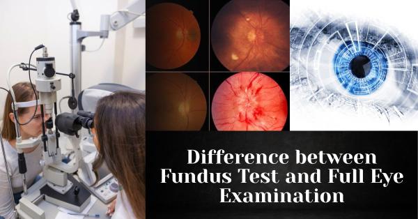
What Is a Fundus Test?
A fundus test is a medical examination that enables eye care specialists to examine the retina, optic nerve, macula, and blood vessels in the back of the eye in order to evaluate its health and identify any possible issues. When pupil dilation is performed to obtain a better vision, it is often referred to as a fundus exam, retinal exam, or dilated eye exam. A light may be shone into the eye while special lenses or cameras are used to take pictures of the internal structures. The process is typically brief, taking only a few minutes. For routine eye health monitoring, managing diabetes or hypertension, checking glaucoma, or when patients report symptoms like visual changes, floaters, or eye pain, doctors usually advise a fundus test.
What Is a Full Eye Examination?
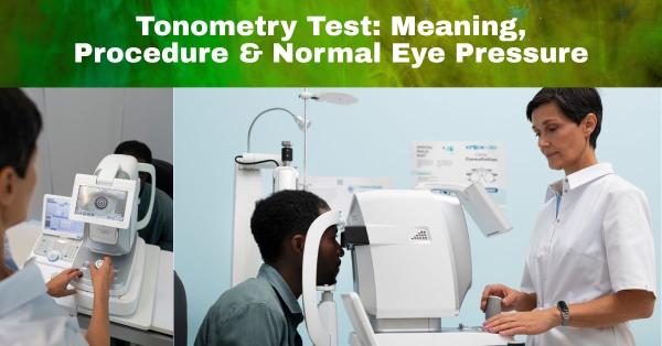
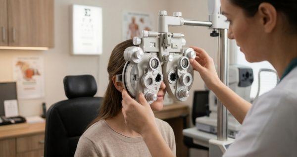
In order to identify vision issues and underlying eye disorders, a full eye examination involves a thorough evaluation of general eye health and visual performance. It addresses every facet of visual health, such as visual acuity, refractive errors, peripheral vision, intraocular pressure, eye muscle function, and the condition of internal and exterior eye structures, unlike a single test. In order to guarantee both clear vision and healthy eyes, a complete eye exam usually consists of tests like a visual acuity test, refraction, slit-lamp examination, fundus (retinal) examination, tonometry for eye pressure, and occasionally additional imaging or color vision assessment.
A comprehensive eye exam includes a number of essential elements, each of which assesses a distinct facet of visual health. The Refraction Test establishes the appropriate prescription for glasses or contact lenses, while the Visual Acuity Test gauges how well you can see at different distances. The cornea, lens, and eyelids are among the front structures of the eye that can be thoroughly examined with the Slit-Lamp Examination. Elevated intraocular pressure, a risk factor for glaucoma, is detected by the Eye Pressure Test (Tonometry). In order to ensure appropriate binocular vision, the Eye Muscle & Movement Test evaluates eye alignment and coordination. A color vision test assesses the capacity to discriminate between various colors in order to detect possible deficits in color vision. In order to identify early indicators of retinal disorders or systemic conditions affecting the eye, the Fundus Examination, which is frequently a part of a comprehensive eye exam, looks at the retina, optic nerve, macula, and blood vessels.
Important Distinctions Between Full Eye Examination and Fundus Test
Test Scope
There are notable differences in the test's scope between the two. The retina, optic nerve, macula, and blood vessels at the rear of the eye are the only internal structures that can be examined with a fundus test. A full eye exam, on the other hand, provides a thorough examination of overall eye health by evaluating the entire eye and visual system, including visual acuity, refractive errors, eye pressure, eye muscles, and both the front and back structures of the eye.
What Each Test Finds
In terms of detection, a fundus test is mostly used to find retinal illnesses such diabetic retinopathy, macular degeneration, changes associated with glaucoma, and other conditions affecting the back of the eye. In contrast, a full eye exam provides a thorough assessment of the entire visual system by identifying refractive errors such as myopia or hyperopia, eye muscle or movement problems, elevated eye pressure, anterior eye problems, color vision deficiencies, and possible retinal or systemic conditions.
Tools and Equipment Used
The two tests employ different tools and equipment. In a fundus test, the retina, optic nerve, macula, and blood vessels in the back of the eye are usually examined with an ophthalmoscope or a fundus camera. A full eye exam, on the other hand, uses a variety of tools to provide a thorough evaluation of the entire eye and visual system. These tools include eye charts for visual acuity, phoropters or autorefractors for refraction, slit-lamp microscopes for anterior eye structures, tonometers for eye pressure, color vision plates, and occasionally imaging devices like OCT scanners.
Duration & Procedure
There are notable differences in the two tests' duration and procedure. Because a fundus test only looks at the retina and adjacent structures, it is typically rapid, taking only a few minutes to complete. A full eye examination, on the other hand, takes longer because it includes a number of tests that provide a comprehensive assessment of overall eye health and visual function, including visual acuity, refraction, slit-lamp inspection, eye pressure measurement, eye muscle testing, color vision evaluation, and a fundus exam.
Dilated vs. Non-Dilated Testing
A fundus test frequently necessitates pupil dilation in order to provide the eye care specialist with a broader and clearer view of the retina, optic nerve, and blood vessels. A complete eye examination, on the other hand, may involve both dilated and non-dilated procedures. Non-dilated tests are used for rapid evaluations such as visual acuity or eye pressure, while dilation is carried out as necessary to fully inspect the internal structures of the eye and identify subtle retinal or optic nerve problems.
Who Should Take Which Test
Depending on personal risk factors and the assessment's goal, different people should take different tests. Since a fundus test focuses on identifying illnesses that impact the back of the eye, it is particularly advised for diabetics, hypertensive patients, and anybody with known or suspected retinal or optic nerve problems. On the other hand, regardless of age or present vision status, a complete eye examination is recommended for everyone as part of normal eye care in order to monitor overall eye health, detect refractive problems, identify early indicators of eye illness, and maintain total visual well-being.
Conditions Found by a Fundus Examination
The purpose of a fundus test is to identify conditions that affect the back of the eye, such as diabetic retinopathy, which damages the blood vessels in the retina; glaucoma, which affects the optic nerve; macular degeneration, which affects central vision; retinal detachment, which is a serious separation of the retina; hypertensive retinopathy, which is brought on by high blood pressure; and other problems with the optic nerve. This test aids in the early detection and treatment of these potentially vision-threatening diseases by looking at the retina, macula, blood vessels, and optic disc.
Conditions Found During a Complete Eye Examination
Numerous visual and eye health problems can be found with a thorough eye exam. It detects refractive defects that impair vision clarity, such as myopia, hyperopia, and astigmatism. Cataracts, dry eyes, squint or alignment abnormalities, and other corneal disorders are frequently detected. The full eye exam is a comprehensive tool for evaluating both overall vision and the structural health of the eyes because it can also identify retinal and optic nerve conditions like diabetic retinopathy, glaucoma, and macular degeneration if a fundus examination is part of the exam.
When Should You Get A Fundus Test?
In certain circumstances where a thorough retinal evaluation is required, you should think about doing a fundus test. It is advised if you have high blood pressure or diabetes because both can impact the blood vessels in your retina. If you have symptoms like floaters, flashes, or abrupt changes in vision, which could be signs of retinal problems, a fundus test is also recommended. Additionally, your eye care specialist can recommend the test as part of yearly retinal monitoring to track current conditions and identify early indicators of disease before symptoms arise, or following a normal eye examination for a closer look at the retina.
When Is a Complete Eye Exam Necessary?
To guarantee complete eye health, a thorough eye exam is advised in a number of circumstances. It should be performed as part of a yearly regular examination for continuous monitoring or if you require glasses or contact lenses to correct your vision. Children benefit from checks as part of school vision screenings to identify problems early, and anyone experiencing changes in their vision or hazy vision should make an appointment for an evaluation right away. Regular examinations may also be necessary for individuals who engage in visually demanding occupations or spend a lot of time on screens in order to avoid eye strain and maintain good visual performance.
In conclusion, a full eye examination uses a variety of eye test instruments, such as visual acuity charts, slit-lamp microscopes, tonometers, and refraction devices, to provide a thorough evaluation of overall vision and eye health, whereas a fundus test uses specialized eye test equipment, such as fundus cameras or ophthalmoscopes, to examine the retina and detect optic nerve conditions. Because early detection of retinal, structural, or refractive problems can avert complications and preserve vision, both exams serve as essential for maintaining overall eye health. Frequent examinations with specialized eye test equipment provide specific diagnosis, prompt treatment, and long-term eye protection.
Author's Bio

Mr. Rajender Gupta
(Director, Matronix Optotechnik Pvt. Ltd.)
With a vision to make advanced eye-care technology accessible across India and beyond, the Director of Matronix Optotechnik Pvt. Ltd. has been leading innovation in smart ophthalmic solutions since founding the company in 2019. Building on decades of industry experience and the global legacy of the Matronix brand since 2007, he has transformed the company into a trusted name in precision eye-testing equipment.


Frequently Asked Questions
A fundus test focuses only on examining the retina, optic nerve, macula, and blood vessels at the back of the eye, usually with pupil dilation. A full eye examination is more comprehensive and evaluates overall eye health and vision, including visual acuity, refractive errors, eye pressure, eye muscle coordination, anterior eye structures, and often includes a fundus examination as part of the process.
Yes, a fundus examination is often included as part of a complete eye exam, especially when the doctor needs to assess retinal health or detect conditions such as diabetic retinopathy, glaucoma, or macular degeneration. However, a fundus test can also be performed separately when a detailed retinal evaluation is required.
A fundus test is recommended for individuals with diabetes, high blood pressure, or a history of retinal or optic nerve disorders. It is also advised for patients experiencing symptoms such as floaters, flashes of light, sudden vision changes, or eye pain, as well as for routine monitoring of existing retinal conditions.
A full eye examination is essential because many eye diseases develop without noticeable symptoms in the early stages. Even if vision appears normal, a comprehensive eye exam can detect refractive errors, increased eye pressure, early cataracts, retinal diseases, and other underlying eye conditions, helping prevent long-term vision loss through early diagnosis and treatment.