
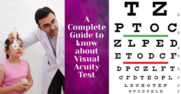
Fundoscopy Test: Purpose, Procedure & Results
Published On: 12/Feb/2026Table of Contents
- • What Is a Fundoscopy Test?
- • Purpose of the Fundoscopy Test
- • Conditions Diagnosed Using Fundoscopy
- • Types of Fundoscopy Tests
- • Fundoscopy Test Procedure
- • What Does a Fundoscopy Test Show?
- • Understanding Fundoscopy Test Results
- • Fundoscopy Test Risks & Safety
- • Fundoscopy Test vs Other Retinal Examinations
- • Who Should Get a Fundoscopy Test?
Fundoscopy, also known as ophthalmoscopy, is an important aspect of a thorough eye exam that enables medical professionals to look for early indications of eye and systemic disorders in the retina, optic nerve, macula, and retinal blood vessels using advanced eye test machines such as a slit lamp with fundus lenses, direct or indirect ophthalmoscopes, and digital fundus cameras. Fundoscopy is essential for quick diagnosis, efficient monitoring, and the prevention of vision loss, as it can identify diseases like diabetic and hypertensive retinopathy, glaucoma, and macular degeneration at an early stage. Frequently advised for individuals with long-term medical conditions, vision or neurological symptoms, and as part of routine eye examinations, fundoscopy supports long-term eye health and overall well-being. Let’s know all about it.
What Is a Fundoscopy Test?
A fundoscopy test, sometimes referred to as an ophthalmoscopy, is a diagnostic eye examination that uses specialized tools or computerized retinal imaging equipment to inspect the back of the eye. It enables eye care specialists to assess the retina, optic disc, macula, and retinal blood vessels all of which are critical for vision and general eye health. Early detection and monitoring of eye conditions such as glaucoma, diabetic retinopathy, macular degeneration, and retinal detachment are made easier by fundoscopy. Additionally, it identifies symptoms of neurological illnesses, diabetes, and hypertension, which can help avoid irreversible eyesight loss with timely treatment.
Purpose of the Fundoscopy Test
The fundoscopy test's objective is to give eye care providers a good view of the back of the eye so they can identify and assess a variety of ocular and systemic diseases early on. Through examination of the retina, fundoscopy assists in the detection of retinal conditions that, if untreated, could endanger eyesight, including diabetic retinopathy, macular degeneration, retinal tears, and infections. In order to identify symptoms of glaucoma, optic nerve enlargement, or atrophy that could point to elevated eye pressure or neurological problems, it is also essential for evaluating the health of the optic nerve. Additionally, because changes in retinal blood vessels frequently reflect general cardiovascular and metabolic health, fundoscopy can identify symptoms of systemic diseases like diabetes, hypertension, and vascular problems. Frequent fundoscopy is also necessary to track the course of the disease and assess the effectiveness of medications, allowing for quick adjustments to care plans and preserving long-term vision.
Conditions Diagnosed Using Fundoscopy
In order to identify serious eye and systemic disorders, fundoscopy is an essential diagnostic procedure that enables direct inspection of the retina and optic nerve. By exposing blood vessel damage brought on by elevated blood pressure or blood sugar, it aids in the diagnosis of diabetic and hypertensive retinopathy. Through distinctive alterations in the optic nerve and macular structure, the test is essential for detecting glaucoma and macular degeneration. Fundoscopy can identify retinal detachment and papilledema in urgent cases, which could be signs of severe neurological disorders.
Types of Fundoscopy Tests
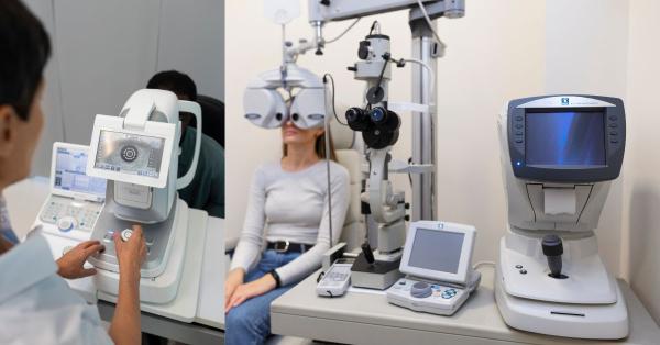
Depending on the therapeutic need, different fundoscopy test types offer varying fields of vision and levels of information. Although technique has a small field of view, direct fundoscopy is helpful for rapid examinations in routine or primary care settings because it uses a handheld ophthalmoscope to provide a magnified, upright image of the central retina and optic disc. Indirect fundoscopy is particularly useful for identifying retinal tears, detachments, and other peripheral retinal abnormalities because it offers a wider, inverted view of the retina and improves visualization of the peripheral retina when done with a handheld lens and a head-mounted light source. Slit-lamp fundoscopy, which is frequently utilized in thorough clinical and specialized eye exams, provides high-resolution, stereoscopic views of the retina and optic nerve by combining a slit-lamp microscope with specific lenses. Advanced fundus cameras are used in digital fundoscopy, commonly referred to as retinal imaging, to provide high-quality digital photographs of the retina. This allows for accurate recordkeeping, simple comparisons across time, telemedicine consultations, and enhanced patient education through visual records.
Fundoscopy Test Procedure
The fundoscopy test is a quick, easy, and typically painless treatment that starts with basic patient preparation, including examining medical history, current medications, and any complaints linked to vision. The patient is instructed to relax and concentrate on a fixed target during the examination. Dilating eye drops are frequently used to dilate the pupils, which enhances retinal and optic nerve visibility. However, this may result in short-term light sensitivity and blurred near vision for a few hours. After the pupils are sufficiently dilated, the eye care specialist examines the retina, macula, optic disc, and retinal blood vessels in a methodical, step-by-step manner using a direct or indirect ophthalmoscope, a slit lamp with special lenses, or a digital fundus camera. The patient is frequently asked to look in different directions to assess both central and peripheral areas. The full fundoscopy examination usually takes 5 to 10 minutes, but if extensive retinal imaging or pupil dilation are needed, it could take a little longer.
What Does a Fundoscopy Test Show?
A fundoscopy test gives medical experts a thorough view of the internal structures in the back of the eye, enabling them to evaluate a number of vital components necessary for good vision. It displays the state of the retina, which transforms light into visual information and aids in the detection of degenerative changes, tears, inflammation, and injury. To diagnose diseases like glaucoma, optic neuritis, or papilledema, the optic disc, the visible part of the optic nerve, is measured for size, shape, color, and cupping. Systemic disorders such as diabetes and hypertension might be indicated by irregular growth, hemorrhages, leakage, or narrowing of the retinal blood vessels. While fundoscopy can identify vitreous abnormalities such floaters, bleeding, or inflammatory changes that could impede clear vision, the macula, which is responsible for sharp center vision, is evaluated for enlargement, degeneration, or deposits that could impair reading and fine detail vision.
Understanding Fundoscopy Test Results
Determining whether the back of the eye is healthy or exhibits symptoms of disease that could impair vision or general health requires an understanding of the fundoscopy test results. A clear macula, a flat, intact retina, a healthy pink optic disc with well-defined borders, and retinal blood vessels of normal size and appearance free of leaks or hemorrhages are all typical fundoscopy findings. Macular changes associated with age-related macular degeneration, retinal hemorrhages or microaneurysms linked to diabetes or hypertension, optic disc cupping or swelling suggestive of glaucoma or elevated intracranial pressure, or retinal tears and detachment that need immediate attention are examples of common abnormal findings. When abnormalities are found or symptoms continue, additional testing is advised. This may involve optical coherence tomography (OCT), visual field testing, fluorescein angiography, or other imaging and laboratory tests to confirm the diagnosis, gauge severity, and direct the right course of treatment or referral to a specialist.
Fundoscopy Test Risks & Safety
The pupil-dilating eye medications used during the examination can cause some temporary side effects, but the fundoscopy test is generally highly safe for people of all ages. These drops often go away in a few hours, although they may cause short-term clouded near vision, light sensitivity, moderate stinging, or difficulty focusing. When carried out by qualified eye care specialists, fundoscopy is regarded as safe for both young patients and senior patients. It is frequently used to identify age-related disorders in older persons and early eye issues in youngsters. Fundoscopy, however, may be avoided or delayed in some circumstances, such as when a patient has a known allergy to dilating drops, is at risk for angle-closure glaucoma, has severe eye pain or trauma that necessitates an immediate alternative assessment, or when acute eye infections or inflammation make the examination uncomfortable or unsafe. In these cases, the test is rescheduled or carried out using non-dilating techniques.
Fundoscopy Test vs Other Retinal Examinations
While retinal photography takes static digital photos that are mostly used for documentation, screening, and follow-up comparisons, fundoscopy is a clinical examination that enables eye care experts to directly observe the retina, optic disc, and blood vessels in real time. Fundoscopy provides a quicker, more comprehensive picture of the general health of the retina, but it has less structural information than optical coherence tomography (OCT), which produces high-resolution cross-sectional images and accurate measurements of retinal layers. Because fundoscopy allows for immediate clinical judgment, dynamic assessment of various retinal areas, and guidance on whether advanced imaging such as OCT or retinal photography is required, it is frequently chosen as the first-line examination during routine eye check-ups, emergency assessments, or when a quick, cost-effective evaluation is required.
Who Should Get a Fundoscopy Test?
A fundoscopy test is advised for a variety of people since it assists in the early detection of systemic and ocular disorders, frequently before symptoms worsen. To track changes in retinal blood vessels and avoid vision-threatening conditions like diabetic or hypertensive retinopathy, people with diabetes or hypertension should have regular fundoscopies. Fundoscopy is necessary to detect potential retinal or optic nerve issues in patients with visual abnormalities, such as impaired vision, flashes, floaters, or abrupt vision loss. Fundoscopy may also be necessary for children with suspected eye or developmental abnormalities in order to rule out neurological, congenital, or retinal conditions that could impair vision. Additionally, fundoscopy is frequently included in adult patients' regular eye exams as a preventive strategy, particularly as they age, to guarantee long-term eye health and facilitate the early diagnosis of diseases like glaucoma and macular degeneration.
A direct and detailed picture of the retina, optic nerve, and blood vessels is provided by fundoscopy, which is an important component of comprehensive eye care. This view serves as essential for evaluating the health of the eye as well as any indications of systemic disease. Its early detection of diseases like diabetic retinopathy, glaucoma, macular degeneration, and neurological illnesses is essential for avoiding long-term problems and irreversible vision loss. Fundoscopy enables fast action, efficient treatment, and improved visual results by detecting issues before obvious symptoms manifest. Because they are essential for preserving good vision, safeguarding general eye health, and guaranteeing early identification of potentially sight-threatening disorders, routine comprehensive eye exams that include fundoscopy should be promoted for individuals of all ages.
Author's Bio

Mr. Rajender Gupta
(Director, Matronix Optotechnik Pvt. Ltd.)
With a vision to make advanced eye-care technology accessible across India and beyond, the Director of Matronix Optotechnik Pvt. Ltd. has been leading innovation in smart ophthalmic solutions since founding the company in 2019. Building on decades of industry experience and the global legacy of the Matronix brand since 2007, he has transformed the company into a trusted name in precision eye-testing equipment.


Frequently Asked Questions
No, a fundoscopy test is usually painless and quick. You may feel mild discomfort from bright lights or experience temporary blurred vision and light sensitivity if pupil-dilating eye drops are used, but these effects typically wear off within a few hours.
The examination itself usually takes 5 to 10 minutes. If pupil dilation or advanced retinal imaging is required, the overall visit may take slightly longer due to the time needed for the eye drops to take effect.
Yes. Fundoscopy can reveal signs of systemic conditions such as diabetes, hypertension, and neurological disorders. Changes in retinal blood vessels or optic nerve swelling often reflect broader health issues, allowing for early diagnosis and timely medical intervention.
The frequency depends on age, medical history, and risk factors. People with diabetes, high blood pressure, glaucoma risk, or vision symptoms may need regular fundoscopies, while others may undergo the test as part of routine comprehensive eye exams for preventive eye care.