VF Test vs OCT – Which Test Is Better for Glaucoma Monitoring?
Published On: 20/Jan/2026Table of Contents
- • What Is a VF Test (Visual Field Test)?
- • What Is OCT (Optical Coherence Tomography)?
- • VF Test vs OCT: Key Differences Explained
- • Which Test Is Better for Tracking Glaucoma Progression?
- • Limitations of VF Test and OCT
- • Do You Need Both VF Test and OCT?
- • How Often Should VF Test and OCT Be Done?
Glaucoma is a collection of progressive eye diseases that harm the optic nerve, frequently as a result of high intraocular pressure. If treatment is not received, glaucoma can cause irreparable vision loss. Regular monitoring is necessary to identify abnormalities early, control development, and maintain vision because the disease usually progresses slowly and might not show any early symptoms. Optical Coherence Tomography (OCT), a procedure that offers detailed imaging of the optic nerve and retinal nerve fiber layer in order to monitor structural damage, and the Visual Field (VF) Test, which evaluates functional vision loss by mapping blind spots and areas of reduced sensitivity, are important tools in glaucoma monitoring. When combined, these tests enable medical personnel to track the anatomical and functional features of glaucoma, assuring immediate care and efficient disease control.
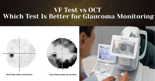
What Is a VF Test (Visual Field Test)?
A vital tool in the treatment of glaucoma, the Visual Field (VF) Test analyzes a person's peripheral and central vision to identify blind spots or areas of vision loss. In order to detect early peripheral vision loss, which frequently happens before central vision is compromised, the test assesses how well the eyes are able to detect things at different positions without moving the head or eyes. A variety of VF tests are employed, such as kinetic perimetry, which monitors moving targets across the visual field, and automated static perimetry, which displays light stimuli at fixed places. The VF test assists eye care providers in tracking the course of glaucoma, evaluating the impact of treatment, and directing prompt measures to stop more vision deterioration by detecting functional vision deficiencies.
What Is OCT (Optical Coherence Tomography)?
A non-invasive imaging technique called optical coherence tomography (OCT) produces high-resolution, cross-sectional images of the retina and optic nerve, enabling medical experts to identify structural alterations linked to glaucoma and other eye disorders. In order to properly visualize the optic nerve head, retinal nerve fiber layer (RNFL), and ganglion cells, OCT uses light waves to take detailed pictures of the various layers of the retina. Even before functional vision loss happens, OCT can detect early structural damage by assessing the thickness and integrity of the RNFL and ganglion cell layer. Because it enables medical professionals to monitor the course of the disease, assess the efficacy of treatment, and act quickly to protect vision, OCT is an essential tool for glaucoma monitoring.
VF Test vs OCT: Key Differences Explained
By evaluating several elements of eye health, the Visual Field (VF) Test and Optical Coherence Tomography (OCT) combine to provide a comprehensive picture of glaucoma. By mapping areas of vision loss or blind patches, the VF test identifies functional impairment and illustrates how glaucoma impacts a patient's actual visual performance. OCT, on the other hand, measures thinning of the retinal nerve fiber layer (RNFL) and ganglion cells even before vision loss is apparent. It does this by taking comprehensive pictures of the optic nerve and retinal layers. Combining the two tests is necessary because structural alterations might occur before functional impairments and vice versa, making it easier for medical personnel to monitor the course of a disease. Together, these advanced eye test equipment solutions enable early detection, improved treatment planning, and more successful prevention of irreversible vision loss in glaucoma patients through complementary VF and OCT data.
Which Test Detects Glaucoma Earlier?
OCT is often more effective at detecting glaucoma in its early stages because it can detect structural alterations in the optic nerve and retinal nerve fiber layer (RNFL) before there is any visible visual loss. OCT enables eye care providers to detect early disease and take immediate action by evaluating ganglion cell destruction and thinning of the RNFL. In contrast, functional vision loss, such as blind spots or decreased sensitivity, is detected by the VF test and usually manifests later in the disease phase. Both techniques, however, have limits in early glaucoma: VF testing may be impacted by patient attention, weariness, or learning effects, thereby missing early functional losses, while OCT may reveal modest changes that are challenging to interpret without baseline data. When combined, OCT and VF offer complementing data that enhances glaucoma development monitoring and early detection.
Which Test Is Better for Tracking Glaucoma Progression?
Both the VF test and OCT serve as essential but complementary tools for monitoring glaucoma development. The VF test is especially useful for long-term functional monitoring since it tracks how a patient's visual field changes over time, illustrating how glaucoma affects day-to-day vision and identifying new blind spots. By measuring the thickness of the optic nerve and retinal nerve fiber layer (RNFL) to detect progressive deterioration before it results in vision loss, OCT, on the other hand, excels in tracking structural changes. For both tests, trend analysis across several visits is essential since isolated readings can be deceptive. Regular monitoring enables doctors to identify minor changes, assess the effectiveness of treatment, and modify management strategies to preserve vision as best they can.
Limitations of VF Test and OCT
Both OCT and the VF test have drawbacks that make them insufficient for managing glaucoma on their own. Accuracy may be impacted by weariness, inattention, or inconsistent responses because the VF test significantly relies on the patient's focus and effort, especially during prolonged or repeated testing sessions. Even though OCT is objective and automated, it may be difficult to interpret due to problems with image quality, media opacities, or typical anatomical changes in the optic nerve and retinal layers. It is important for using both VF and OCT together to acquire a complete, accurate picture of glaucoma status since each test evaluates different parts of the illness, functional versus structural, and relying just on one test may overlook early changes or subtle development.
Do You Need Both VF Test and OCT?
For thorough glaucoma management, both the VF test and OCT are usually advised because they offer complimentary information about the condition. In clinical practice, physicians are using the VF test to track functional vision loss over time and OCT to identify early anatomical alterations in the optic nerve and retinal nerve fiber layer (RNFL). The stage of glaucoma, patient risk factors, and previous test results determine which test to prioritize and how often to do each. Together, VF and OCT provide a comprehensive evaluation of both structural and functional characteristics, enabling precise diagnosis, early management, and efficient tracking of disease development. Relying just on one method may overlook subtle progression or early alterations, which is why combined testing is significant.
How Often Should VF Test and OCT Be Done?
Whether an individual is being screened for glaucoma or has already been diagnosed with the condition determines how frequently VF and OCT tests are performed. Every one to two years, eye care specialists may advise a thorough examination that includes OCT and VF testing for general screening in at-risk patients, such as older adults or those with a family history of glaucoma. The frequency of monitoring for individuals with glaucoma depends on the severity of the condition and how quickly it is progressing. For example, patients with early or stable glaucoma may need testing every six to twelve months, while those with advanced or rapidly growing glaucoma may require more frequent evaluations, sometimes every three to six months. In order to ensure quick evaluation of changes and efficient management of glaucoma throughout time, testing strategies are customized, accounting for variables such as intraocular pressure, optic nerve appearance, previous test results, and overall risk.
In the treatment of glaucoma, the VF test and OCT have distinct but complementary functions. While OCT offers thorough structural imaging of the optic nerve and retinal nerve fiber layer to detect early anatomical changes, the VF test detects functional vision loss by identifying blind spots and decreased sensitivity in the visual field. Because glaucoma can impact both structure and function at different stages, combining these tests provides the most accurate evaluation, enabling eye care providers to identify the condition early, track its progression, and successfully customize therapy. In order to protect your vision and preserve long-term eye health, a combined approach promotes the significance of routine eye exams and makes sure that both structural and functional changes are monitored over time.
Author's Bio

Mr. Rajender Gupta
(Director, Matronix Optotechnik Pvt. Ltd.)
With a vision to make advanced eye-care technology accessible across India and beyond, the Director of Matronix Optotechnik Pvt. Ltd. has been leading innovation in smart ophthalmic solutions since founding the company in 2019. Building on decades of industry experience and the global legacy of the Matronix brand since 2007, he has transformed the company into a trusted name in precision eye-testing equipment.


Frequently Asked Questions
The Visual Field (VF) test measures functional vision loss by identifying blind spots and areas of reduced sensitivity, showing how glaucoma affects a patient’s visual performance. OCT, on the other hand, detects structural damage by imaging the optic nerve and retinal nerve fiber layer (RNFL), often identifying changes before noticeable vision loss occurs.
OCT generally detects glaucoma earlier because it can identify structural changes in the optic nerve and RNFL before functional vision loss develops. VF testing usually reveals damage at a later stage, once visual field defects become measurable.
No, relying on only one test may miss early or subtle disease changes. VF and OCT provide complementary information—functional and structural—so using both together offers a more accurate and complete assessment of glaucoma progression.
The testing frequency depends on disease severity and progression. Patients with stable or early glaucoma may need VF and OCT testing every 6–12 months, while those with advanced or rapidly progressing glaucoma may require testing every 3–6 months, as determined by their eye care specialist.