Confrontation Testing: Meaning, Procedure and Uses
Published On: 22/Feb/2026Table of Contents
- • What Is Confrontation Testing?
- • Meaning of Confrontation Testing
- • Purpose of Confrontation Testing
- • Conditions Detected Using Confrontation Testing
- • Types of Confrontation Testing
- • Confrontation Testing Procedure
- • What Confrontation Testing Evaluates
- • Understanding Confrontation Test Results
- • Accuracy & Limitations of Confrontation Testing
- • Confrontation Testing vs Automated Perimeter
- • Uses of Confrontation Testing in Clinical Practice
Visual field testing is an important aspect of a comprehensive eye examination since it assesses the entire range of what a person can see, including central and peripheral side vision. Peripheral vision is essential for mobility, spatial awareness, and movement detection, while central vision enables us to read and focus on fine details. As such, its evaluation is critical for detecting conditions like glaucoma, retinal disorders, and neurological diseases that may result in gradual or abrupt vision loss. Among the several approaches, confronting visual field testing is a straightforward, efficient, and reasonably priced screening tool that is frequently used to identify evident visual field abnormalities during standard eye and neurological examinations. This simple test assists doctors in detecting possible anomalies early and determining whether more sophisticated visual field testing is necessary by comparing the examiner's visual field with the patient's vision.
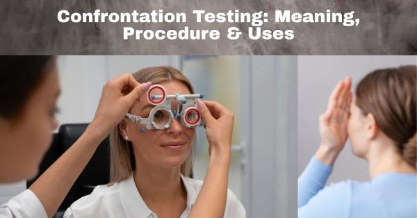
What Is Confrontation Testing?
Confrontation testing is a simple bedside technique for evaluating the visual field in which the examiner presents fingers or visual targets in various regions of the field of view in order to compare the patient's peripheral vision to their own. This visual field assessment test's main goal is to promptly identify severe visual field abnormalities, like hemianopia or peripheral vision loss, which may be linked to diseases like glaucoma, stroke, or other neurological problems. Contrary to automated visual field tests like computerized perimetry, which employ specialized equipment to map visual sensitivity in detail and quantify subtle defects, confrontation testing is primarily used as a quick screening tool during routine eye and neurological examinations rather than for precise diagnosis or monitoring. It also requires no instruments and only provides a general estimate of the visual field.
Meaning of Confrontation Testing
The capacity to see objects and movement outside of one's direct line of sight is known as peripheral side vision, and it is essential for balance, spatial awareness, and safe everyday navigation. Visual field defects, which can be caused by neurological issues like stroke or brain tumors, or eye diseases like glaucoma or retinal disease, are areas of this entire visual field that are absent or diminished.Confrontation testing is used as a quick and easy screening method during routine eye and neurological exams to detect obvious peripheral vision loss because these defects may not always be immediately apparent to patients. This helps clinicians identify potential abnormalities early and determine whether more sophisticated visual field testing is required for a thorough evaluation.
Purpose of Confrontation Testing
Confrontation visual field testing is frequently used to screen for visual field loss because it enables medical professionals to promptly spot important peripheral vision gaps in patients that could otherwise go overlooked. Healthcare professionals can identify possible neurological abnormalities like stroke, brain tumors, or optic pathway lesions, as well as ocular problems like glaucoma or retinal disorders, by evaluating a patient's ability to perceive visual stimuli in various locations of their field of view. Its ease of use, rapidity, and lack of equipment requirements make it particularly useful in emergency situations and routine clinical examinations, where a quick evaluation of visual function can yield essential hints for prompt diagnosis and treatment.
Conditions Detected Using Confrontation Testing
Confrontation testing can aid in the detection of a variety of serious ocular and neurological diseases by detecting major vision field abnormalities during regular examinations. Notable field constriction can be used to detect growing peripheral vision loss in disorders like glaucoma early on. Blind patches or partial field loss are common symptoms of optic nerve illnesses, such as optic neuritis or compressive optic neuropathy. While retinal conditions like retinal detachment or advanced diabetic retinopathy can also cause localized or extensive vision gaps, sudden visual field deficits may indicate stroke or brain tumors impacting the visual pathways. Confrontation testing is a useful first screening method for identifying potentially dangerous underlying issues since different neurological disorders that affect the brain's visual processing centers may manifest with distinct patterns of vision field loss.
Types of Confrontation Testing
A number of straightforward methods can be used for confrontation testing in order to assess various visual field components. The finger counting technique helps determine the boundaries of the patient's gross visual field by having the examiner hold out one or more fingers in different peripheral positions and ask the patient to count how many they saw. In order to identify more serious field loss, the hand movement detection method involves the patient indicating when they notice movement in their side vision. The object comparison approach looks for asymmetries by comparing the examiner's visual field with the patient's ability to perceive a small target. The red target confrontation test, which can be especially useful in detecting anomalies of the optic nerve, evaluates color vision and central field problems using a red object.
Confrontation Testing Procedure
In order to ensure that only one eye is examined at a time, confrontation testing involves the patient being seated comfortably at eye level with the examiner, usually one meter apart, in a well-lit room. The patient is then told to focus on the examiner's open eye while covering the other eye. After closing the matching eye, the examiner shows the patient fingers or a visual target in various quadrants of their peripheral field. The patient is then asked to count the number of fingers displayed or to indicate when they perceive the item. To compare the patient's reactions with the examiner's own visual field as a reference, this methodical procedure is repeated in each of the four visual field quadrants. The test is then conducted on the opposite eye. It is easy to administer and doesn't require any special equipment, so it normally just takes a few minutes to finish. This makes it perfect for rapid screening in both routine and emergency situations.
What Confrontation Testing Evaluates
Confrontation testing helps detect any narrowing or loss of the visual field by measuring how far into the side fields a patient can detect visual stimuli. This allows for an evaluation of the overall extent of peripheral vision. In order to identify unequal or asymmetric field abnormalities that can indicate localized ocular or neurological issues, it also evaluates the symmetry between the two eyes by comparing responses from the right and left sides. The test is a useful first screening tool in clinical practice because it also helps identify blind spots or visual field defects, such as partial or complete areas of missing vision, which can be linked to conditions affecting the retina, optic nerve, or visual pathways in the brain.
Understanding Confrontation Test Results
The first step in comprehending confrontation test results is realizing that normal findings demonstrate complete and symmetrical peripheral vision in both eyes' quadrants, with the patient detecting visual stimuli simultaneously and in the same locations as the examiner. Common aberrant patterns include localized blind spots (scotomas), loss of vision in one half of the visual field (hemianopia), or diminished peripheral vision (field constriction). Such abnormal results usually call for additional evaluation with more thorough visual field testing and imaging studies to confirm the diagnosis. These abnormal results may indicate underlying conditions such as glaucoma, retinal disease, optic nerve damage, stroke, brain tumors, or other neurological disorders affecting the visual pathways.
Accuracy & Limitations of Confrontation Testing
The Humphrey Visual Field Analyzer and other automated perimeter tests are more accurate than confrontation visual field testing, which is a quick and practical bedside screening procedure. Its sensitivity for early or subtle abnormalities is generally low, therefore minor scotomas or moderate glaucomatous alterations may go undetected, even though it can detect big or dense visual field defects (such as hemianopia or severe glaucoma). Examiner experience, patient compliance, attention span, weariness, poor fixation, and irregular testing distance or lighting conditions are some of the variables that can impact reliability. Due to these limitations, advanced automatic perimeter is advised when glaucoma is suspected, when neurological conditions (such as stroke, brain tumors, or optic nerve disorders) are being assessed, when symptoms continue despite normal confrontation results, or when accurate documentation and progression monitoring are necessary.
Confrontation Testing vs Automated Perimeter
Unlike automated perimetry and other advanced eye test instruments, confrontation testing does not require specialized equipment and is primarily used as a rapid screening tool rather than a diagnostic method.. The sensitivity and accuracy of automated perimetry, which offers comprehensive, quantitative maps of the visual field and detects early or subtle defects like those seen in glaucoma, are superior to confrontation testing, a quick, easy, bedside screening technique that can detect large or obvious field defects. In primary care settings, emergency evaluations, immobile patients, or situations requiring rapid neurological screening without specialist equipment, confrontation testing is recommended. On the other hand, because automated perimetry enables precise documenting and tracking of progression, it is important for the diagnosis and monitoring of problems such as glaucoma, optic nerve diseases, and neurological disorders. Confrontation testing and automated perimeter work together as a screening technique, and automated perimeter is essential for long-term disease management and comprehensive eye care.
Uses of Confrontation Testing in Clinical Practice
In clinical practice, confrontation testing is frequently employed as a rapid and useful technique to evaluate the visual field in diverse contexts. It assists in identifying clear visual field abnormalities that could be a sign of glaucoma or issues with the optic nerve during routine eye exams. Finding patterns of vision loss, like hemianopia or quadrantanopia, which could indicate diseases like stroke, brain tumors, or other intracranial lesions, is necessary in neurological evaluations. Because it doesn't require specific equipment and can be done right away on critically ill or immobilized patients, it is particularly useful in emergency and bedside evaluations. Confrontation testing is an accessible first-line evaluation in both ophthalmology and general medical practice, and it is also an effective screening technique in primary care settings to identify patients who require referral for further visual field testing.
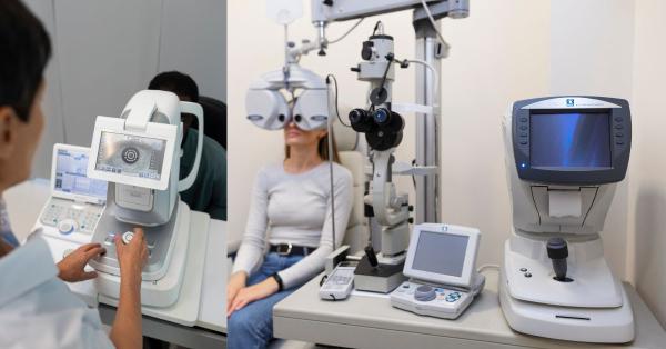
Particularly in regular examinations, neurological evaluations, and primary care settings, confrontation testing is a useful and significant screening tool that aids in the early diagnosis of visual field abnormalities. It can promptly detect major field losses like hemianopia or advanced glaucomatous damage, allowing for immediate referral and care, even if it is less sensitive than automated perimetry. It is particularly helpful for the early suspicion of neurological or optic nerve problems because of its affordability, ease of usage, and lack of specialist equipment. Following an abnormal confrontation test, a comprehensive ocular examination using a Slit Lamp is often essential to assess the anterior segment, optic nerve head, and other ocular structures that may contribute to visual field defects. Although confrontation testing is affordable, simple to perform, and requires no specialized equipment, subtle or early abnormalities may be missed; therefore, abnormal findings should always be followed by detailed visual field testing and clinical examination to confirm the diagnosis, assess severity, and guide appropriate management for optimal long-term visual outcomes.
Author's Bio

Mr. Rajender Gupta
(Director, Matronix Optotechnik Pvt. Ltd.)
With a vision to make advanced eye-care technology accessible across India and beyond, the Director of Matronix Optotechnik Pvt. Ltd. has been leading innovation in smart ophthalmic solutions since founding the company in 2019. Building on decades of industry experience and the global legacy of the Matronix brand since 2007, he has transformed the company into a trusted name in precision eye-testing equipment.

Frequently Asked Questions
Confrontation testing is a simple bedside visual field assessment used to screen for obvious peripheral vision loss by comparing the patient’s visual field with that of the examiner. It is performed during routine eye or neurological examinations to quickly identify major visual field defects that may indicate conditions such as glaucoma, stroke, or optic nerve disorders.
Confrontation testing can help detect significant visual field abnormalities associated with glaucoma, optic nerve diseases (like optic neuritis), retinal conditions, stroke, brain tumors, and other neurological disorders that affect the visual pathways, especially when defects are large or advanced.
Confrontation testing is a quick, equipment-free screening tool used to identify gross visual field defects, while automated perimetry provides detailed, quantitative mapping of the visual field and can detect subtle or early changes. Automated perimetry is essential for diagnosis, monitoring disease progression, and long-term management, whereas confrontation testing is mainly for initial screening.
Although confrontation testing is simple and cost-effective, it has low sensitivity for detecting early or subtle visual field defects. Results can also be influenced by examiner experience, patient cooperation, and testing conditions. Therefore, any abnormal or suspicious findings should be followed by comprehensive eye examinations and automated visual field testing for accurate diagnosis and management.