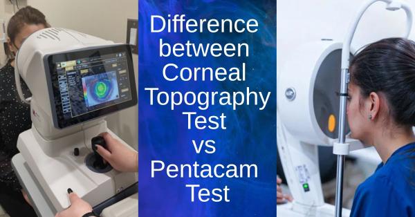
Difference between Pentacam vs Corneal Topography
Published On: 29/Mar/2026Corneal imaging is important in modern eye care because it provides extensive information about the shape, thickness, and overall health of the cornea, allowing for accurate diagnosis and treatment planning. Planning refractive surgeries like LASIK, identifying disorders like keratoconus, and guaranteeing safe results in procedures like cataract surgery all depend on accurate corneal assessment. The Pentacam and corneal topography are two of the most popular tools for this purpose; both provide insightful information on the structure and curvature of the cornea, assisting medical professionals in making well-informed therapeutic decisions.
What Is Corneal Topography?
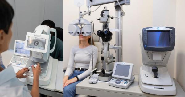
A non-invasive diagnostic method called corneal topography helps medical professionals comprehend the shape and optical characteristics of the cornea by producing a thorough map of its surface curvature. It detects even minute curvature anomalies by projecting concentric rings onto the cornea (usually utilizing Placido disc-based systems) and examining the reflected patterns. In clinical practice, this technology is frequently used to assess candidates for refractive operations like LASIK, fit contact lenses, diagnose disorders like keratoconus, and track changes in corneal shape over time. Such capabilities highlight how modern eye test machines support accurate corneal evaluation and improve overall diagnostic efficiency in routine eye care.
What Is Pentacam?
The Pentacam is a modern diagnostic tool used in eye care that offers a thorough and in-depth examination of the anterior portion of the eye. Considered one of the most advanced eye test instruments, Its foundation is Scheimpflug imaging technology, which produces an accurate three-dimensional (3D) model by taking many cross-sectional photographs of the eye. In contrast to simple surface mapping methods, the Pentacam gathers comprehensive information on the anterior chamber and lens, including corneal thickness, elevation, and curvature, in addition to evaluating the cornea. Because of this, it is particularly useful for planning refractive and cataract procedures, identifying corneal problems, and guaranteeing precise and secure clinical results.
Key Differences Between Pentacam and Corneal Topography
Technology Featured: The Pentacam uses a rotating Scheimpflug camera to take multiple cross-sectional images, enabling detailed 3D imaging of the anterior and posterior corneal areas as well as deeper eye parts, whereas corneal topography uses Placido disc reflection to evaluate the curvature of the cornea's front surface by interpreting reflected ring patterns.
Area of Analysis: The Pentacam provides a more thorough evaluation by examining both the anterior and posterior corneal surfaces in addition to the anterior chamber, providing a deeper and more comprehensive understanding of eye structure, whereas corneal topography only assesses the anterior (front) surface of the cornea, primarily providing information about surface curvature.
Data Results: The Pentacam provides more detailed information, such as elevation maps, corneal thickness (pachymetry), anterior chamber depth, and detailed 3D analysis of the anterior segment for a more thorough clinical evaluation, whereas corneal topography mainly provides curvature maps that depict the shape and steepness of the cornea's front surface.
Accuracy and Specifications: Corneal topography is more constrained because it primarily focuses on the anterior surface, but it is still a helpful and popular tool for basic corneal assessment. In contrast, the Pentacam offers more thorough and accurate data by analyzing both anterior and posterior corneal surfaces along with additional parameters.
Detection of Corneal Conditions: Corneal topography is primarily useful for identifying surface irregularities on the front of the cornea, making it useful for initial screening and routine evaluations, whereas Pentacam is more effective for early detection of conditions like keratoconus because it analyzes both anterior and posterior corneal changes along with thickness variations.
Clinical Applications
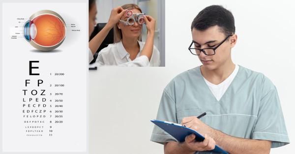
In routine clinical practice, corneal topography is frequently used to assess the form and curvature of the cornea's front surface. By assisting practitioners in choosing lenses that complement the corneal contour for the best comfort and vision, it plays a significant part in contact lens fitting. It is also helpful in detecting simple corneal imperfections that could impair vision quality and screening for refractive defects like astigmatism. Corneal topography is frequently the first-line method for routine corneal examination and initial screening because of its ease of use and effectiveness.
The Pentacam, on the other hand, is employed in more sophisticated and intricate clinical applications.As part of advanced eye test equipment, it offers thorough information on corneal thickness, elevation, and structure to guarantee patient safety and the best possible results, it is essential for planning refractive procedures like LASIK and PRK. By evaluating the anterior chamber and lens position, the Pentacam is also very useful in the diagnosis and tracking of keratoconus, particularly in its early stages, and is important in the assessment of cataract surgery. It is a useful instrument for accurate diagnosis and long-term treatment of a variety of eye problems due to its capacity to measure corneal thickness (pachymetry) and offer a comprehensive 3D analysis.
Which One Is Better?
The particular clinical necessity and the degree of detail required for diagnosis or therapy planning ultimately determine which approach is superior. Because it offers a thorough three-dimensional analysis of the anterior segment, including anterior and posterior corneal surfaces, corneal thickness, and anterior chamber parameters, the Pentacam is typically regarded as superior for advanced diagnostics. This makes it extremely useful for early detection of conditions like keratoconus and for immediate planning of procedures like LASIK, PRK, and cataract surgery. It has a distinct benefit in complicated or high-risk situations because of its capacity to identify minute structural alterations that would not be apparent on surface-based examinations. Corneal topography, on the other hand, continues to be a vital and useful tool in routine eye care because of its speed, affordability, and accessibility. It provides accurate information about the cornea's front surface for tasks like contact lens fitting, astigmatism screening, and detecting simple surface irregularities. In many clinical contexts, both technologies are utilized in tandem rather than in place of one another, with Pentacam providing deeper analysis when a more thorough assessment is needed and corneal topography acting as an initial screening tool to ensure correct diagnosis and the best possible patient treatment.
When Are Both Used Together?
Corneal topography and the Pentacam are frequently used in conjunction in complex or borderline cases requiring a more thorough evaluation of the cornea, as they provide complementing information. The Pentacam provides deeper insights into posterior corneal alterations, thickness distribution, and anterior chamber details, whilst topography provides a fast evaluation of the anterior surface curvature. Combining the information from both instruments improves diagnostic precision, aids in the identification of subtle or early corneal abnormalities, and facilitates improved decision-making regarding treatments like refractive surgery or the management of corneal disorders.
In summary, Pentacam and corneal topography both have significant but different functions in corneal evaluation; Pentacam offers a more thorough 3D study of the entire anterior segment, whereas topography concentrates on anterior surface curvature. The clinical need determines which diagnostic tool is best since employing the proper technology guarantees exact diagnosis, efficient treatment planning, and improved eye condition monitoring. Ultimately, by facilitating early diagnosis, accurate assessment, and safer surgical outcomes, the use of sophisticated imaging systems like Pentacam, either by itself or in conjunction with corneal topography, greatly improves patient outcomes.
Author's Bio

Mr. Rajender Gupta
(Director, Matronix Optotechnik Pvt. Ltd.)
With a vision to make advanced eye-care technology accessible across India and beyond, the Director of Matronix Optotechnik Pvt. Ltd. has been leading innovation in smart ophthalmic solutions since founding the company in 2019. Building on decades of industry experience and the global legacy of the Matronix brand since 2007, he has transformed the company into a trusted name in precision eye-testing equipment.

Frequently Asked Questions
The main difference is that corneal topography analyzes only the front surface of the cornea, while Pentacam provides a detailed 3D analysis of both the anterior and posterior corneal surfaces along with additional parameters like corneal thickness and anterior chamber depth.
Pentacam is generally considered more accurate for advanced diagnosis because it evaluates both front and back surfaces of the cornea and provides comprehensive data. Corneal topography is reliable for basic assessments but is limited to surface-level analysis.
Yes, Pentacam can detect keratoconus at an earlier stage because it analyzes posterior corneal changes and thickness variations, which are not captured by standard corneal topography.
Both are used together in complex or borderline cases where detailed corneal evaluation is required. Topography provides quick surface mapping, while Pentacam adds deeper structural insights, improving diagnosis and treatment planning.