How OCT Is Revolutionizing Diabetic Retinopathy Screening in India
Published On: 31/May/2026Table of Contents
- • The Diabetic Retinopathy Crisis in India
- • Traditional Retinal Screening Methods & Their Limitations
- • Understanding OCT: What It Is and How It Works
- • How OCT Is Transforming Diabetic Retinopathy Screening
- • Early Detection of Diabetic Macular Edema (DME)
- • Accurate Staging of Diabetic Retinopathy
- • Monitoring Treatment Response
- • Faster & Higher Patient Throughput
- • Telemedicine & Remote Screening
- • OCT vs Other Retinal Imaging Methods
- • Real-World Impact: OCT in Indian Healthcare Settings
- • What to Look for When Buying an OCT for Your Clinic
- • Axial Resolution
- • Scan Speed
- • Scan Width and Depth Coverage
- • Built-in Retinal Thickness Mapping and Analysis Software
- • Ease of Use for Technicians
- • Export and Telemedicine Compatibility
- • After-Sales Service and Calibration Support in India
- • Price Suitability for Indian Clinic/Hospital Budgets
- • The Road Ahead: AI + OCT in Diabetic Retinopathy
India has over 101 million people living with diabetes, yet most will never receive a retinal exam. Diabetic retinopathy, one of the leading causes of preventable blindness in working-age adults, is advancing silently in millions of eyes right now. There is a need for quicker and more effective diagnostic options because traditional retinal screening techniques are frequently slow, specialist-dependent, and challenging to access in smaller towns.
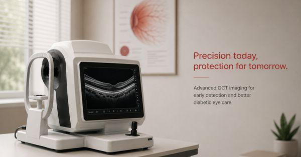
By offering immediate, non-invasive, and incredibly detailed retinal imaging that promotes early identification and improved disease management, Optical Coherence Tomography (OCT) is revolutionizing diabetic eye care. Clinics and hospitals around India are increasingly using OCT for routine diabetic retinopathy screening as it becomes more accessible and economical.
The Diabetic Retinopathy Crisis in India
As one of the world's major centers for diabetes patients, India is still dealing with a developing diabetic retinopathy (DR) crisis, which has resulted in a concerning increase in diabetes-related eye disorders. According to studies, almost one in three people with diabetes may get diabetic retinopathy at some point in their lives, therefore routine retinal screening is important to managing the condition. However, a significant issue is that the majority of DR cases go untreated until patients start to noticeably impair their vision, at which point retinal degeneration may have progressed and become more challenging to treat.
In tier-2 and tier-3 cities and rural areas, where access to trained ophthalmologists, retinal imaging equipment, and organized screening programs is still restricted, the issue is particularly problematic. As a result, many individuals either receive delayed diagnoses or undergo no eye exams at all. In order to maintain vision and prevent irreversible blindness, early detection is vital because diabetic retinopathy is mostly preventable and treatable when detected in its early stages. This allows for timely therapies such as medication, laser therapy, lifestyle management, or injections.
Traditional Retinal Screening Methods & Their Limitations
For many years, traditional retinal screening techniques have been fundamental to the treatment of diabetic eye disease, but each has serious limitations that might prevent early identification and disease tracking. Fundus photography produces two-dimensional surface images of the retina and is helpful for recording obvious errors in the retina, but it is unable to show modest fluid buildup beneath the surface or changes in the deeper retinal layer. Ophthalmologists can visually inspect the retina in real time using direct and indirect ophthalmoscopy, but both techniques lack a permanent digital record that can be compared over time, and their accuracy is largely dependent on the examiner's skill and experience. Fundus Fluorescein Angiography (FFA) is an invasive treatment that needs intravenous dye injection, can be time-consuming, and may cause discomfort or allergic responses in certain individuals, despite being a highly effective method for assessing retinal blood vessel leakage and circulation problems. Common diagnostic gaps exist in all of these traditional methods, such as the inability to identify diabetic macular edema in its early stages, the lack of high-resolution cross-sectional retinal imaging, and the absence of accurate quantitative data for tracking the course of the illness. The early detection and treatment of diabetic retinopathy are much improved by the thorough, non-invasive, and extremely accurate retinal imaging that Optical Coherence Tomography (OCT) provides.
Understanding OCT: What It Is and How It Works
In ophthalmology, optical coherence tomography (OCT) is a highly sophisticated non-invasive imaging technique that produces clear, high-resolution cross-sectional images of the retina, enabling medical experts to analyze its tiny structure with remarkable clarity. Similar to ultrasound imaging, the technology employs light waves to scan the eye. However, OCT uses reflected light rather than sound waves to create exact retinal images. This makes it possible for medical professionals to see individual retinal layers at micron-level resolution, allowing them to identify even minute retinal abnormalities that might not be apparent using traditional inspection techniques.
One of the main benefits of OCT is that there is no dye injection, no physical contact with the eye, and no radiation exposure, making the treatment entirely safe and comfortable for patients. OCT is a vital tool for the early diagnosis, monitoring, and treatment of diabetic retinopathy and other retinal problems since the scan itself is rapid, painless, and extremely effective, with detailed data usually available within seconds.
Near-infrared light is directed into the eye toward the retina. Depending on the structure and density of each retinal layer, the light is reflected back in a different way. Highly sensitive sensors pick up these reflected light signals, which are then processed by sophisticated software algorithms to produce intricate cross-sectional images that are frequently referred to as "optical slices" of the retina. This enables medical professionals to see the retina layer by layer with amazing accuracy and evaluate even small structural flaws that might not be apparent during a standard examination.
These high-resolution scans allow eye care specialists to accurately measure retinal thickness, detect fluid buildup, and identify early indicators of diabetic macular edema. Structural changes can also be tracked over time with precision, enabling better long-term disease management. With several advanced tools able to scan the entire macular region in less than two seconds, modern OCT systems are incredibly swift and efficient. This makes the process quick, pleasant, and very useful for routine diabetic retinopathy screening and follow-up therapy.
How OCT Is Transforming Diabetic Retinopathy Screening
By providing quick, non-invasive, and extremely detailed retinal imaging that aids in the early detection of retinal damage and diabetic macular edema, optical coherence tomography (OCT) is revolutionizing the screening process for diabetic retinopathy. Its capacity to offer cross-sectional, quantitative examination of retinal layers enhances diagnostic precision, facilitates swift intervention choices, and increases the effectiveness and accessibility of advanced diabetic eye care.
Early Detection of Diabetic Macular Edema (DME)
Early identification is necessary for maintaining vision because diabetic macular edema (DME) is one of the main causes of vision loss in patients with diabetic retinopathy. By generating extremely accurate cross-sectional retinal pictures, OCT enables medical professionals to detect even preclinical macular edema before patients have obvious visual issues.
In order to make quick and effective treatment decisions, its retinal thickness mapping capability additionally assists in measuring fluid accumulation and exact tracking edema progression over time.
Accurate Staging of Diabetic Retinopathy
By offering a detailed visualization of retinal structures, optical coherence tomography (OCT) enables highly accurate staging of diabetic retinopathy, assisting clinicians in correctly classifying the condition into mild, moderate, severe non-proliferative diabetic retinopathy (NPDR) or proliferative diabetic retinopathy (PDR).
It can easily identify deformities that may be challenging to evaluate using traditional diagnostic techniques, such as intraretinal fluid, subretinal fluid, and epiretinal membranes. OCT considerably lessens the diagnostic uncertainty frequently connected with conventional ophthalmoscopy by providing objective and high-resolution images.
Monitoring Treatment Response
By providing reliable, consistent retinal measurements, OCT is essential for tracking the beneficial effects of diabetic retinopathy treatments like anti-VEGF injections and laser therapy.
Clinicians can monitor changes in retinal thickness and fluid buildup over time with serial OCT scans, which can help assess whether the treatment is effectively lowering edema and enhancing retinal health. Instead of depending only on subjective clinical evaluation, this allows for more specific, data-driven therapy recommendations.
Faster & Higher Patient Throughput
By enabling quick and efficient retinal imaging, OCT greatly increases workflow efficiency in diabetic eye screening. A full macular scan, including patient preparation, usually takes less than five minutes. Many contemporary OCT systems, especially wide-field OCT devices, can conduct scans without the need for pupil dilation, making the process more convenient and comfortable for patients. Hospitals, eye clinics, and high-volume diabetic centers can screen more patients daily while retaining excellent diagnostic accuracy because of its speed and simplicity of use.
Telemedicine & Remote Screening
By enabling high-resolution retinal pictures to be digitally preserved and shared with retinal specialists for expert review from any place, OCT technology significantly supports telemedicine and remote diabetic retinopathy screening.
This enables efficient eye screening camps to be held in underdeveloped and rural parts of India where local retinal specialists might not be available. OCT improves early detection and immediate assistance for diabetic patients across larger regions by facilitating remote diagnosis and consultation, thereby bridging the gap between urban and rural eye care access.
OCT vs Other Retinal Imaging Methods
By delivering high-resolution cross-sectional retinal pictures, early identification of retinal edema, and quantitative study of retinal thickness, optical coherence tomography (OCT) has several advantages over conventional retinal imaging techniques. OCT is quick, non-invasive, and able to see individual retinal layers in great detail, in contrast to fundus photography, ophthalmoscopy, or FFA. To show the distinctions, the table below contrasts OCT with other widely used retinal imaging methods.
.
|
Feature |
OCT |
Fundus Camera |
FFA |
|
Invasive? |
No |
No |
Yes (Dye Injection) |
|
Cross Sectional View |
Yes |
No |
No |
|
Detects macular edema |
Yes (early) |
Limited |
Yes (late stage) |
|
Patient comfort |
High |
High |
Low |
|
Time per scan |
2-5 mins |
5-10 mins |
20-30 mins |
|
Quantitative data |
Yes |
No |
Partial |
|
Ideal for monitoring |
Yes |
No |
No |
Real-World Impact: OCT in Indian Healthcare Settings
With top institutes like AIIMS, Sankara Nethralaya, and LV Prasad Eye Institute increasingly incorporating modern OCT technology into regular retinal diagnoses and diabetic retinopathy management, optical coherence tomography (OCT) is quickly revolutionizing diabetic eye care throughout India. Additionally, OCT has grown to be an essential component of national diabetic retinopathy screening programs, assisting medical professionals in early detection of retinal impairment and improving the course of therapy for diabetic patients.
The use of OCT systems is spreading beyond metropolitan hospitals into tier-2 cities and developing healthcare facilities as they become more reasonably priced and technologically sophisticated, increasing access to specialized retinal imaging for a larger population. OCT is also being used to facilitate widespread diabetic eye screening in underprivileged communities through government healthcare programs and NGO-led eye screening camps, allowing for quicker diagnosis and early referrals.
At Matronix, we supply affordable optical coherence tomography oct designed specifically for the Indian healthcare environment from high-volume city hospitals to emerging tier-2 and tier-3 clinics. If you're evaluating OCT for your practice, speak with our team to find the right fit.
What to Look for When Buying an OCT for Your Clinic
As OCT adoption grows across Indian healthcare settings, choosing the right eye test machines for your clinic is critical. Here are the key factors to evaluate before making a purchase decision.
Axial Resolution
The OCT system's ability to see individual retinal layers and identify minute irregularities depends on its axial resolution. It is expressed in microns, and a lower micron number denotes improved diagnostic accuracy and crisper image quality, particularly for early diabetic retinal alterations.
Scan Speed
The number of A-scans the OCT device can complete in a second is referred to as scan speed. Increased scan speeds minimize examination times, enhance picture quality, lessen motion artifacts, and assist clinics in effectively handling larger patient volumes.
Scan Width and Depth Coverage
Clinicians can view deeper retinal structures and capture bigger retinal areas in a single scan thanks to a wider and deeper scan range. This is especially helpful for detecting peripheral retinal alterations and conducting a thorough evaluation of diabetic retinopathy.
Built-in Retinal Thickness Mapping and Analysis Software
Automated retinal thickness maps and analysis tools that assist in measuring edema, comparing changes over time, and producing objective diagnostic reports are features of advanced OCT systems. These characteristics streamline illness monitoring and enhance clinical decision-making.
Ease of Use for Technicians
Technicians with less experience may operate the device more easily thanks to its user-friendly software, simple controls, and automated alignment functions, which lessen operator dependency. This guarantees constant scan quality and increases workflow efficiency.
Export and Telemedicine Compatibility
Digital image export, cloud storage, and telemedicine integration for remote consultations and expert evaluation should be supported by contemporary OCT instruments. This is particularly useful for remote screening programs and multi-center clinics.
After-Sales Service and Calibration Support in India
Maintaining device performance and reducing downtime requires consistent calibration support, technical help, and dependable after-sales service. Clinics want to select vendors who have a strong service network throughout India.
Price Suitability for Indian Clinic/Hospital Budgets
Depending on the number of patients and diagnostic requirements of the clinic, the OCT system should provide the ideal balance between innovative characteristics and affordability. For expanding practices and tier-2 or tier-3 healthcare facilities, affordable solutions with long-term dependability are essential.
The Road Ahead: AI + OCT in Diabetic Retinopathy
Artificial Intelligence (AI) and Optical Coherence Tomography (OCT) are progressively influencing the future of diabetic retinopathy screening, opening the door to more rapid, intelligent, and easily accessible retinal diagnostics. With increasing accuracy, sophisticated AI algorithms are currently being trained to automatically evaluate OCT scans and identify indicators of diabetic retinopathy, diabetic macular edema, and other retinal abnormalities. Particularly in areas with a shortage of retinal specialists, this technique could greatly increase screening efficiency in community clinics and basic health centers.
Researchers and medical facilities in India are also creating India-specific retinal imaging datasets that more accurately reflect the anatomical and pathological patterns observed in Indian patients in order to improve diagnostic accuracy for local populations. The country's rising diabetes burden may be more successfully addressed if OCT and AI are combined to enable widespread diabetic eye screening programs with less expert intervention. In order to enable the next generation of intelligent eye care solutions, forward-thinking makers of ophthalmic equipment are increasingly concentrating on cloud connectivity, automated analytics, telemedicine integration, and AI-ready OCT systems.
By offering fast, accurate, non-invasive retinal imaging that promotes early identification and improved treatment of diabetic eye disease, optical coherence tomography (OCT) is revolutionizing diabetic retinopathy screening. Clinics and hospitals in India are using it more and more to enhance patient care while getting ready for the increased need for sophisticated retinal diagnostics. With businesses like Matronix providing reasonably priced OCT systems designed for Indian healthcare settings, investing in OCT technology allows healthcare professionals to improve screening efficiency, protect patient vision, and future-proof their practice.
Author's Bio

Mr. Rajender Gupta
(Director, Matronix Optotechnik Pvt. Ltd.)
With a vision to make advanced eye-care technology accessible across India and beyond, the Director of Matronix Optotechnik Pvt. Ltd. has been leading innovation in smart ophthalmic solutions since founding the company in 2019. Building on decades of industry experience and the global legacy of the Matronix brand since 2007, he has transformed the company into a trusted name in precision eye-testing equipment.

Frequently Asked Questions
Optical Coherence Tomography (OCT) is a non-invasive imaging technology that uses light waves to create detailed cross-sectional images of the retina. It helps detect diabetic retinopathy and diabetic macular edema (DME) at an early stage by revealing subtle retinal changes that may not be visible through conventional eye examinations.
Unlike fundus photography and ophthalmoscopy, OCT provides high-resolution cross-sectional views of retinal layers and offers quantitative measurements of retinal thickness. It can detect fluid buildup and early signs of diabetic macular edema more accurately, enabling earlier diagnosis and timely treatment.
Yes. One of the major advantages of OCT is its ability to identify retinal swelling, fluid accumulation, and structural changes before noticeable vision problems develop. Early detection allows ophthalmologists to begin treatment sooner and reduce the risk of permanent vision loss.
Clinics should evaluate factors such as axial resolution, scan speed, scan depth and width coverage, retinal thickness analysis software, ease of use, telemedicine compatibility, after-sales service support, and overall affordability. Selecting the right OCT system helps improve diagnostic accuracy, workflow efficiency, and patient care outcomes.