High-Speed Optical Coherence Tomography System
Product Specification :-
| Vertical Resolution | ≤5μm |
| Horizontal Resolution | ≤20μm |
| Field of View | 40° H x 40° V |
| Anesthesia Requirement | Required |
| Minimum Pupil Diameter | 2mm |
| Refractive Adjustment Range | -18D to +16 D |
| Scan Speed | 250,000 a Scan/second |
| Imagining Technology | Confocal Scaning Laser Ophthalmoscopy |
| Optic Disc | Retinal Nerve Fiber Layer Thickness; Cup-to-disc Ration; Binocular Contrast |
| Retinal Vessel Segmentation | Retina Full Layer Retina Superficial Layer; Retina Deep Layer; Retina Avascular Laye; Choroid Layer |
| Scaning Mode | Scanning positions include macular region; optic disc region; and anterior segment; Scanning range options include 3mmx3mm, 6mmx6mm; Scanning Density320*320*2A-Scan |
| Artificial Intelligence Auxiliary diagnostic function | Optional open network port, to achieve single-line high-definition scanning online Al intelligent auxiliary diagnosis function |
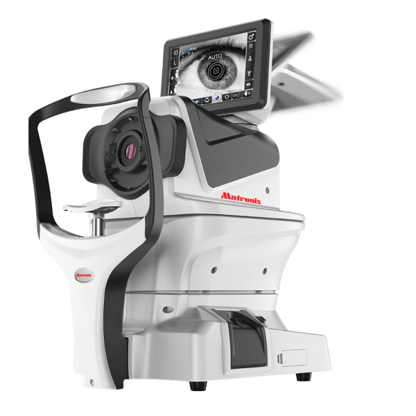
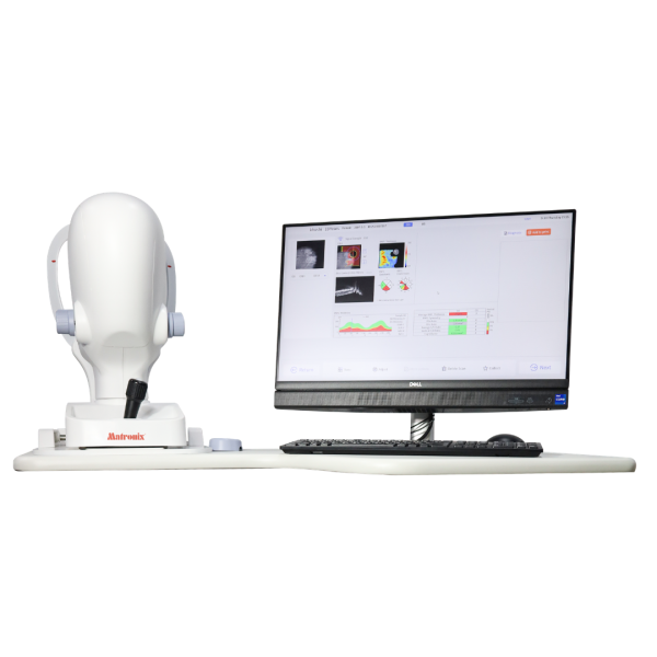
High-Speed Optical Coherence Tomography System for Advanced Retinal Imaging
The Matronix Optical Coherence Tomography (OCT) System is an advanced diagnostic imaging device designed to deliver high-definition cross-sectional images of retinal and anterior segment structures. Built with high-speed scanning technology and AI-assisted diagnostic tools, the system enables eye care professionals to perform detailed analysis of retinal layers, optic nerve structures, and anterior chamber angles with exceptional precision.
With a scan speed of up to 250,000 A-scans per second, the Matronix OCT provides ultra-fast imaging while maintaining excellent resolution for detailed tissue visualization. The device captures high-definition scans of the macula, optic disc, and anterior segment, supporting comprehensive ophthalmic examinations and disease monitoring.
Powered by an 840 nm light source, the system achieves vertical resolution ≤5 μm and horizontal resolution ≤20 μm, enabling clinicians to observe microscopic retinal structures with remarkable clarity. The OCT also integrates AI auxiliary diagnosis, offering intelligent analysis support that helps improve clinical efficiency and diagnostic accuracy.
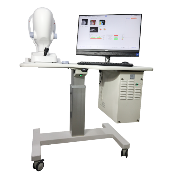
Designed for modern ophthalmology practices, the Matronix OCT provides comprehensive scanning modes, advanced analysis software, and OCT angiography capabilities, making it suitable for glaucoma evaluation, retinal disease diagnosis, and anterior segment analysis.
Key Features
🔹 Ultra-High Speed Scanning
Captures up to 250,000 A-scans per second for fast and efficient imaging.
🔹 High-Resolution Imaging
Provides ≤5 μm vertical resolution for detailed visualization of retinal structures.
🔹 AI-Assisted Diagnosis
Intelligent diagnostic support helps improve clinical decision-making.
🔹 Multi-Region Scan Capability
Supports scanning of macula, optic disc, and anterior segment structures.
🔹 Ganglion Cell Analysis (GCA)
Detects early glaucoma damage through detailed ganglion cell layer analysis.
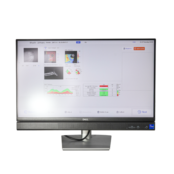
🔹 Optic Disc & RNFL Analysis
Measures retinal nerve fiber layer thickness and cup-to-disc ratio for glaucoma evaluation.
🔹 OCT Angiography (OCTA)
Enables non-invasive visualization of retinal blood vessels.
🔹 Gonioscopy Imaging & Measurement
Allows anterior chamber angle analysis to assist in diagnosing open-angle or closed-angle glaucoma.
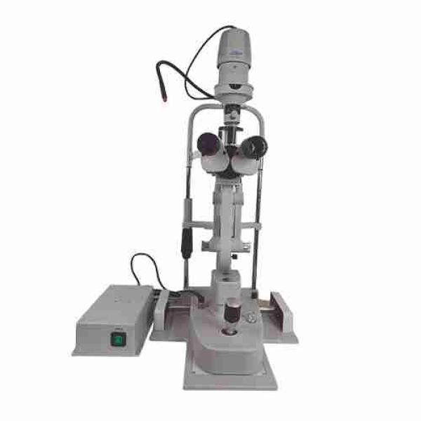
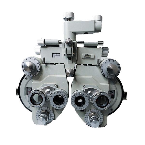
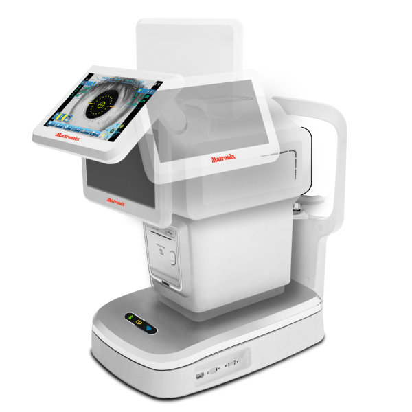
Related Products