Slit Lamp Exam - A Complete Guide for Eye Care Professionals
Published On: 17/Jun/2026Table of Contents
- • What Is a Slit Lamp Examination?
- • Why is the Slit Lamp Examination done?
- • How is the Slit Lamp Examination performed?
- • Patient Preparation
- • Examination Process
- • Use of Fluorescein Dye
- • What Are the Risks of a Slit Lamp Exam?
- • What Are the Illumination Techniques Used in a Slit Lamp Exam?
- • Diffuse Illumination
- • Direct Focal Illumination
- • Optical Section Technique
- • Specular Reflection
- • Retroillumination
- • Sclerotic Scatter
- • What Is Examined During a Slit Lamp Test?
- • Anterior Segment Examination
- • Posterior Segment Examination
- • What are the Results of Slit Lamp Examination?
- • Which Conditions Are Diagnosed with a Slit Lamp Examination?
- • Corneal Disorders
- • Lens Disorders
- • Glaucoma-Related Findings
- • Retinal Conditions
- • Ocular Surface Diseases
- • What Are the Benefits of a Slit Lamp Examination?
- • What are the Limitations of a Slit Lamp Examination?
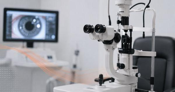
One of the most fundamental diagnostic techniques in eye care is a slit lamp examination, which enables medical professionals to closely inspect the eye's anatomy under specialized light and extreme magnification.
What Is a Slit Lamp Examination?
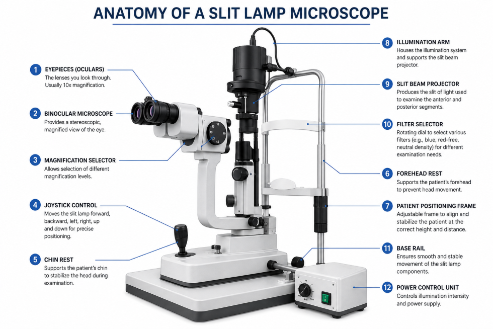
A common eye test that enables eye care specialists to thoroughly examine the front and rear of the eye is the slit lamp examination. A slit lamp microscope, which combines a strong microscope with a bright, adjustable light beam that can be focused on particular parts of the eye, is used for the procedure. This makes it easier for medical professionals to see anatomical features such the cornea, iris, lens, retina, and eyelids in order to identify anomalies, injuries, or diseases of the eyes.
A binocular microscope for magnified visual inspection, a slit-shaped illumination system that offers focused light, a chin rest and forehead support to keep the patient's head steady, and a variety of lenses or attachments that assist in examining various parts of the eye are the main parts of a slit lamp machine. A key element of thorough eye exams and disease identification is this non-invasive, painless test.
Why is the Slit Lamp Examination done?
A slit lamp examination allows eye care experts to closely inspect the eye and detect any signs of disease, injury, or abnormalities that would not be seen during an ordinary vision test.
Doctors can identify eye diseases including cataracts, glaucoma, corneal infections, dry eye disease, and retinal abnormalities early on, frequently before symptoms become apparent, because of the high magnification and concentrated illumination.
Additionally, it is used to measure changes in a patient's general eye health over time, assess recovery following eye surgery or injury, and track the progress of continuing therapy. The slit lamp examination is essential for thorough diagnosis, immediate treatment, and long-term vision care because it offers a comprehensive view of the eye's structure.
How is the Slit Lamp Examination performed?
Eye care specialists can quickly and painlessly examine various areas of the eye under focused light and high magnification with a slit lamp examination. The examination is carried out methodically to evaluate eye health, identify anomalies, and accurately diagnose a range of eye disorders.
Patient Preparation
The patient is instructed to sit comfortably in front of the slit lamp equipment, lay their forehead against the support bar, and rest their chin on the chin rest before the examination starts.
To guarantee a clear view of the eye structures, the eye care specialist then modifies the slit lamp's height, focus, and light settings.
An efficient and comfortable examination is made possible by proper placement, which also keeps the head steady.
Examination Process
The eye care specialist uses a powerful light beam and high magnification to carefully check various regions of the eye during the slit lamp examination.
An external evaluation of the eyelids, lashes, and surrounding tissues typically starts the examination.
After assessing the cornea for scratches, infections, or other anomalies, the anterior chamber is examined to look for indications of inflammation or elevated intraocular pressure.
Then the lens is examined for any abnormalities that could impair vision, such as cataracts. In order to diagnose problems like diabetic retinopathy, glaucoma, and retinal illnesses, special auxiliary lenses are used when necessary to check the retina, macula, and optic nerve at the back of the eye.
Use of Fluorescein Dye
The doctor may inject a tiny quantity of fluorescein dye into the eye during a slit lamp examination.
When exposed to a blue light, this harmless orange dye briefly brightens the surface of the eye. It assists the physician in identifying corneal abrasions, scratches, ulcers, and other corneal damage that can be hard to spot otherwise.
By seeing how uniformly the tears spread across the eye and how quickly they break up, fluorescein dye is also helpful for assessing the tear film, enabling the physician to diagnose dry eye issues and other irregularities.
What Are the Risks of a Slit Lamp Exam?
A slit lamp examination has relatively few side effects and is a non-invasive, painless, and safe treatment. The majority of persons have no negative side effects either during or after the test.
Dilating eye drops may cause light sensitivity and momentary blurred vision for a few hours.
Rarely, the eye drops may cause minor irritation or an allergic reaction in certain individuals.
All things considered, a slit lamp exam is a common, low-risk diagnostic procedure performed in eye clinics all over the world.
What Are the Illumination Techniques Used in a Slit Lamp Exam?
Given below are the techniques used for slit lamp exam:
Diffuse Illumination
This technique uses a broad beam of light to provide an overall view of the eye's external and anterior structures.
Direct Focal Illumination
It focuses a narrow beam directly on a specific area to examine details and abnormalities.
Optical Section Technique
This creates a thin cross-sectional view of the cornea or lens to assess their depth and layers.
Specular Reflection
It uses reflected light to evaluate the health and smoothness of the corneal endothelium and tear film.
Retroillumination
It illuminates eye structures from behind to highlight opacities, defects, or abnormalities.
Sclerotic Scatter
This one directs light through the cornea to detect corneal scars, edema, or other subtle changes.
What Is Examined During a Slit Lamp Test?
Using magnification and concentrated light, a slit lamp test enables eye care specialists to closely inspect the front and rear structures of the eye. It aids in assessing the condition of the cornea, iris, lens, retina, optic nerve, conjunctiva, eyelids, and other significant ocular structures.
Anterior Segment Examination
The front structures of the eye, such as the conjunctiva, cornea, anterior chamber, iris, lens, and eyelids and lashes, are assessed using the slit light during the anterior segment examination.
Infections, inflammation, corneal injuries, dry eye disease, cataracts, and other anomalies that could impair vision and general eye health can all be found with the use of this thorough evaluation.
Posterior Segment Examination
The posterior segment examination uses a slit lamp and specific lenses to evaluate the retina, macula, optic nerve, and vitreous humor in the back of the eye.
Diabetic retinopathy, age-related macular degeneration, retinal tears, glaucoma-related optic nerve damage, and other retinal illnesses that can impair vision are among the conditions that eye care specialists can identify and track using this test.
What are the Results of Slit Lamp Examination?
A slit lamp examination, unlike many other medical examinations, yields no numerical data or a positive/negative outcome.
Rather, the eye care specialist thoroughly inspects your eye's structures and discusses any anomalies, issues, or conditions that might need more testing or care. They will outline the next stages and available treatments if any problems are found.
Which Conditions Are Diagnosed with a Slit Lamp Examination?
Following are the conditions diagnosed by this exam
Corneal Disorders
Corneal abrasions, keratitis, and corneal ulcers are just a few of the conditions that can be diagnosed with a slit lamp examination.
Keratitis is an inflammation of the cornea brought on by an infection, trauma, or extended usage of contact lenses, whereas corneal ulcers are open sores on the cornea that are frequently caused by infections or traumas.
Scratches on the ocular surface known as corneal abrasions can result in pain, redness, and light sensitivity.
The slit lamp enables eye care specialists to accurately identify these conditions and choose the best course of action by using high magnification and specialized illumination.
Lens Disorders
A slit lamp examination is commonly used to diagnose cataracts, a condition in which the eye's natural lens becomes cloudy and affects vision. The high magnification provided by the slit lamp allows eye care professionals to closely examine the lens, identify the presence of cataracts, and determine their type and severity.
Early detection through a slit lamp exam helps in planning timely treatment and preventing further vision impairment.
Glaucoma-Related Findings
Early detection of glaucoma, a disorder brought on by elevated intraocular pressure that can eventually harm the visual nerve, is greatly aided by a slit lamp examination.
The eye care specialist looks for pigment deposits or pseudoexfoliation material on the lens and cornea that are linked to specific types of glaucoma, assesses the depth and angle of the anterior chamber to check for narrow-angle or angle-closure glaucoma, and examines the cornea for signs of swelling associated with elevated eye pressure.
Retinal Conditions
When combined with specialist diagnostic lenses, a slit lamp examination can assist in identifying and tracking a number of retinal disorders, such as age-related macular degeneration (AMD) and diabetic retinopathy.
It enables eye care specialists to look for indications of blood vessel damage, hemorrhage, swelling, or degeneration that could impair vision in the retina and macula. For timely care and to avoid visual loss, early detection of these disorders is necessary.
Ocular Surface Diseases
Ocular surface conditions like conjunctivitis and dry eye syndrome are frequently diagnosed using a slit lamp examination. It assists medical professionals in looking for indications of dryness, irritation, redness, inflammation, or infection on the corneal surface, conjunctiva, and tear film.
This thorough assessment helps in accurate diagnosis and assists in directing the right course of action to enhance eye comfort and preserve good vision.
What Are the Benefits of a Slit Lamp Examination?
A slit lamp examination has several key advantages in eye care since it is a non-invasive, painless method that provides a highly detailed image of the eye's structures.
In order to make an accurate diagnosis, eye care specialists can closely examine the cornea, lens, retina, and other ocular tissues thanks to its strong magnification and specialized illumination.
The examination aids in the early detection of eye illnesses such glaucoma, cataracts, corneal disorders, and retinal disorders, frequently before serious symptoms appear.
A slit lamp exam facilitates better treatment planning, continuous monitoring, and better patient outcomes by detecting issues early on and offering comprehensive clinical information.
What are the Limitations of a Slit Lamp Examination?
A slit lamp examination has several limitations despite being an important eye test machine for eye disorders. The expertise and experience of the eye care specialist conducting the test have a significant impact on its accuracy.
Certain retinal diseases may need advanced imaging procedures like OCT or fundus photography for a more thorough evaluation, and some structures at the rear of the eye may not be fully visible without extra lenses or pupil dilation.
Additionally, the examination may occasionally be more challenging due to patient mobility, poor cooperation, or acute eye irritation. To ensure a thorough assessment of eye health, slit lamp results are often paired with additional diagnostic testing.
Author's Bio

Mr. Rajender Gupta
(Director, Matronix Optotechnik Pvt. Ltd.)
With a vision to make advanced eye-care technology accessible across India and beyond, the Director of Matronix Optotechnik Pvt. Ltd. has been leading innovation in smart ophthalmic solutions since founding the company in 2019. Building on decades of industry experience and the global legacy of the Matronix brand since 2007, he has transformed the company into a trusted name in precision eye-testing equipment.
Related Posts


Frequently Asked Questions
A normal slit lamp examination takes between 5 and 15 minutes, depending on the aim of the test and whether pupil dilation is necessary.
No, a slit lamp examination is entirely non-invasive and painless. The inspection light may only momentarily shine on the patient.
Under normal examination conditions, scrapes, ulcers, corneal injuries, and anomalies in the tear film might not be apparent, but fluorescein dye helps highlight them.
The process is really safe. Dilating drops may cause temporary blurry vision or light sensitivity in certain patients.
Age, eye health, and risk factors all affect the frequency. It is regularly carried out during standard eye exams and more frequently for patients who already have eye disorders.