
What Is Ophthalmoscopy? A Complete Guide for Eye Care Professionals
Published On: 14/Jul/2026Table of Contents
- • What Is Ophthalmoscopy?
- • What are the types of Ophthalmoscopy?
- • Direct Ophthalmoscopy
- • Indirect Ophthalmoscopy
- • Slit Lamp Ophthalmoscopy
- • Why Is Ophthalmoscopy Important?
- • How Is an Ophthalmoscopy Performed?
- • What is Examined During Ophthalmoscopy?
- • Retina
- • Optic Disc
- • Macula
- • Retinal Blood Vessels
- • Vitreous Humor
- • What are the risks of Ophthalmoscopy?
- • What Can Be Diagnosed with Ophthalmoscopy?
- • Diabetic Retinopathy
- • Glaucoma
- • Age-Related Macular Degeneration (AMD)
- • Retinal Detachment
- • Hypertensive Retinopathy
- • Optic Neuritis
- • Papilledema
- • Retinal Vein or Artery Occlusion
- • What are the limitations of Ophthalmoscopy?
Ophthalmoscopy is an important eye exam that is used in present-day eye care to assess the retina, optic nerve, and other interior eye tissues. It helps in the early detection of serious vision conditions such diabetic retinopathy, glaucoma, retinal detachment, and age-related macular degeneration. You will discover what ophthalmoscopy is, how it is done, its types, advantages, and the disorders it can be used to diagnose below.
What Is Ophthalmoscopy?
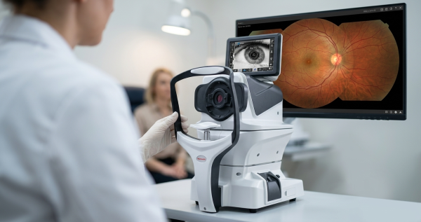
Ophthalmoscopy, sometimes referred to as fundoscopy or a fundus examination, is a diagnostic eye test that enables eye test professionals to look at the retina, optic nerve, macula, and retinal blood vessels at the rear of the eye. The process helps identify anomalies that might not be apparent during a standard eye exam by projecting light into the eye using a specialized tool known as an ophthalmoscope, which provides a clear picture of these interior structures.
Ophthalmoscopy is an important component of comprehensive eye exams because it allows for the early detection and monitoring of vision-threatening conditions such as diabetic retinopathy, glaucoma, age-related macular degeneration, and retinal detachment, resulting in more timely treatment and better results for patients.
What are the types of Ophthalmoscopy?
Ophthalmoscopy is carried out utilizing a variety of methods, each intended to offer a particular perspective of the eye's interior structures. The area being inspected with an eye test machine, the patient's health, and the degree of information needed for a correct diagnosis all influence the method selection.
Direct Ophthalmoscopy
It is carried out with a handheld direct ophthalmoscope, which provides an upright, enlarged image of the retina and optic disc by shining a light beam into the eye. It is perfect for routine eye exams and basic retinal assessments in clinics and hospitals because it is rapid, portable, and simple to use. However, it has a narrow field of view and poor peripheral retinal visualization, particularly in the absence of pupil dilation. During routine eye exams, direct ophthalmoscopy is frequently used to identify anomalies of the optic nerve, diabetic retinopathy, hypertensive retinopathy, and other disorders affecting the central retina.
Indirect Ophthalmoscopy
This is done to get a wide-angle view of the retina using a portable condensing lens and a head-mounted light source. It is particularly helpful for identifying retinal tears and detachments because it offers a wider field of vision than direct ophthalmoscopy, enabling clinicians to study the central and peripheral retina in more detail. Ophthalmology clinics, emergency eye exams, the diagnosis of retinal diseases, the screening for diabetic retinopathy, and the assessment of patients with ocular injuries or other complicated retinal problems frequently opt for this method.
Slit Lamp Ophthalmoscopy
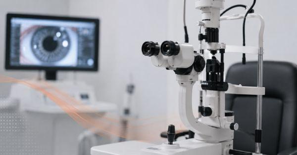
It involves utilizing a slit lamp biomicroscope and a specific fundus lens to obtain a greatly magnified, detailed picture of the retina, optic nerve, and macula. Because of its superior image clarity and depth, eye care specialists can more accurately identify tiny retinal abnormalities. This method is frequently used in eye clinics and hospitals to do thorough retinal examinations, diagnose glaucoma, diabetic retinopathy, macular disorders, and track the development of various eye conditions.
Why Is Ophthalmoscopy Important?
Since it enables eye care specialists to identify retinal and optic nerve abnormalities before they result in obvious vision issues, ophthalmoscopy is a key diagnostic test. In order to enable timely medical care and improved patient outcomes, it is essential for the early detection of illnesses such diabetic retinopathy, glaucoma, age-related macular degeneration, retinal detachment, and other vision-threatening diseases.
Ophthalmoscopy helps monitor systemic illnesses like diabetes and hypertension that might impact the eyes, in addition to assessing the health of the retina and optic nerve. Through routine monitoring and follow-up, the examination helps lower the risk of irreversible vision loss, improves preventive eye care, and directs treatment options by spotting early symptoms of damage.
How Is an Ophthalmoscopy Performed?
The patient's medical history and visual complaints are reviewed by the eye care specialist before the examination. A clearer and more detailed image of the retina, optic nerve, and other internal structures of the eye can be obtained by using dilating eye drops to dilate the pupils, depending on the objective of the test. A slit lamp with a fundus lens, an indirect ophthalmoscope, or a direct ophthalmoscope are then used for the examination.
Although patients may experience momentary bright lights during the test, the treatment usually takes five to fifteen minutes and is painless. Blurred vision and heightened light sensitivity may last for several hours if pupil dilation is utilized. The specialist evaluates the eye, discusses the results, and, if abnormalities are found, suggests further testing or therapy.
What is Examined During Ophthalmoscopy?
The eye care specialist carefully inspects the internal structures at the back of the eye during an ophthalmoscopy in order to evaluate their condition and spot any abnormalities. This thorough assessment assists in the early detection of conditions affecting the retina, optic nerve, and blood vessels that could impair vision.
Retina
The retina is inspected for indications of injury, tears, hemorrhage, swelling, or conditions including age-related macular degeneration and diabetic retinopathy. Clear eyesight and light detection depend on a functioning retina.
Optic Disc
To determine the condition of the optic nerve, the optic disc is examined. Conditions including glaucoma, visual neuritis, or papilledema may be detected by changes like swelling, cupping, or pallor.
Macula
The macula is examined for anomalies such as macular degeneration, macular edema, or other retinal conditions that could impact central vision. It is in charge of the fine, detailed vision required for face recognition and reading.
Retinal Blood Vessels
Narrowing, bleeding, blockages, and other vascular alterations are assessed in the retinal blood vessels. These results can be used to identify systemic disorders like diabetes and hypertension as well as eye problems.
Vitreous Humor
Floaters, hemorrhage, inflammation, and other anomalies that can point to retinal tears, infections, or vitreous diseases are examined in the vitreous humor, the transparent gel that fills the inside of the eye.
What are the risks of Ophthalmoscopy?
Ophthalmoscopy is a low-risk, painless, non-invasive, and harmless eye exam. Patients may have temporary adverse effects, such as minor stinging during application, impaired vision, and increased sensitivity to bright light, while using dilating eye drops to enlarge the pupils. These side effects are common and typically go away a few hours after the test.
Ophthalmoscopy hardly ever results in serious consequences. Rarely, some people may develop a transient rise in ocular pressure or an allergic reaction to the dilating drops, especially if they are at risk for angle-closure glaucoma. Following the treatment, patients who experience acute eye pain, chronic blurred vision, redness, or abrupt changes in vision should seek medical attention right away.
What Can Be Diagnosed with Ophthalmoscopy?
Ophthalmoscopy offers a thorough picture of the retina, optic nerve, and retinal blood vessels, which aids in the diagnosis of a variety of eye disorders. Additionally, it can identify symptoms of systemic conditions affecting the eyes, allowing for timely diagnosis and treatment.
Diabetic Retinopathy
Diabetes-related retinal blood vessel damage, such as microaneurysms, hemorrhage, and edema, can be identified by ophthalmoscopy. Early diagnosis and immediate intervention can help avoid vision loss.
Glaucoma
The examination looks for indications of cupping or glaucoma-related damage to the optic nerve. Frequent monitoring assists in early disease detection and progression management.
Age-Related Macular Degeneration (AMD)
AMD-related abnormalities in the macula, such as drusen deposits or retinal injury, can be detected by ophthalmoscopy. Central vision is preserved with early detection.
Retinal Detachment
The process enables the eye care specialist to detect retinal tears or detachment, which need to be treated immediately to avoid irreversible vision loss.
Hypertensive Retinopathy
Ophthalmoscopy can reveal changes in the retinal blood vessels brought on by high blood pressure, such as swelling, hemorrhage, or narrowing. The degree of hypertension may be indicated by these results.
Optic Neuritis
Ophthalmoscopy can identify optic nerve inflammation or swelling, which is frequently linked to neurological disorders like multiple sclerosis and can result in abrupt vision loss.
Papilledema
Ophthalmoscopy can identify optic disc swelling brought on by elevated intracranial pressure. To identify the underlying reason for this observation, a medical evaluation must be conducted right away.
Retinal Vein or Artery Occlusion
By displaying irregular blood flow, retinal hemorrhages, or areas of diminished blood supply, ophthalmoscopy contributes to the early detection and treatment of blockages in the retinal veins or arteries.
What are the limitations of Ophthalmoscopy?
Despite being a vital diagnostic tool, ophthalmoscopy has limitations. Without pupil dilation, the retina's view may be limited, making a comprehensive examination of the peripheral retina challenging.
Additionally, because subtle retinal or optic nerve abnormalities might be difficult to detect, the accuracy of the examination depends on the experience and competence of the eye care specialist. For a more thorough assessment, indirect ophthalmoscopy may be necessary for certain peripheral retinal diseases, such as retinal tears or detachments.
Media opacities, like cataracts, corneal scarring, or vitreous hemorrhage, can further lower the quality of the examination by making the retina less visible.
Additionally, additional diagnostic tests like Optical Coherence Tomography (OCT), fundus photography, fluorescein angiography, or visual field testing may be required to confirm the diagnosis and direct the proper course of treatment because ophthalmoscopy alone may not be able to provide enough information for all eye conditions.
Author's Bio

Mr. Rajender Gupta
(Director, Matronix Optotechnik Pvt. Ltd.)
With a vision to make advanced eye-care technology accessible across India and beyond, the Director of Matronix Optotechnik Pvt. Ltd. has been leading innovation in smart ophthalmic solutions since founding the company in 2019. Building on decades of industry experience and the global legacy of the Matronix brand since 2007, he has transformed the company into a trusted name in precision eye-testing equipment.
Related Posts

Frequently Asked Questions
No, ophthalmoscopy is a painless and non-invasive eye examination. While bright lights may cause temporary discomfort and dilating eye drops can blur vision for a few hours, the procedure itself does not cause pain.
The frequency depends on your age, eye health, and medical history. People with diabetes, glaucoma, high blood pressure, or a family history of eye disease may need regular ophthalmoscopy as recommended by their eye care professional.
Yes. In addition to diagnosing retinal and optic nerve conditions, ophthalmoscopy can reveal signs of systemic diseases such as diabetes, hypertension, and increased intracranial pressure, which may affect the blood vessels and optic nerve in the eye.
Ophthalmoscopy is performed using specialized eye test machines such as a direct ophthalmoscope, indirect ophthalmoscope, or a slit lamp biomicroscope with a fundus lens. These instruments help eye care professionals examine the retina, optic nerve, and other internal eye structures in detail.